
Advanced Dental Technology Near You
Dental technology has seen remarkable advancements in recent years, making dental appointments faster and more comprehensive. Many of the more time-consuming tasks in dentistry have been simplified, improving efficiency and effectiveness in various procedures.
As technology continues to transform our daily lives at home and at work, it was only a matter of time before these innovations began reshaping how patients experience routine dental visits. Here are some of the cutting-edge technologies we use in our office to enhance your care.
-
Digital X-Rays
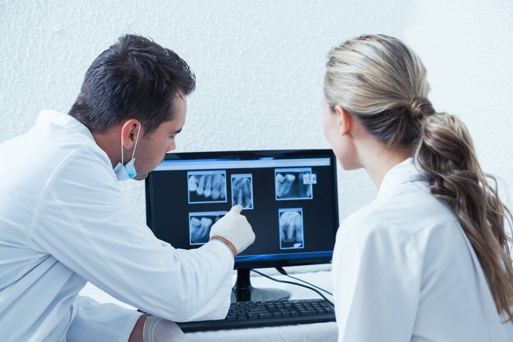
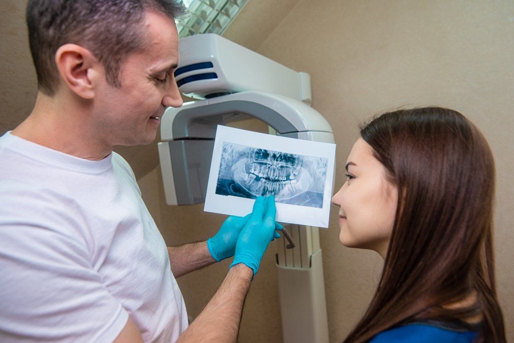
Introduced in 1987, nearly 90 years after traditional x-rays came to fruition, digital radiography combined the power of computer technology with electric sensors and tiny bursts of radiation. Rather than printing the results on film, images form almost as soon as the sensors are placed in our mouths, projecting on a computer screen. Digital x-ray technology does demand additional training for dentists, though the majority of practitioners are adamant that the advantages are worth the commitment. Today, a lot of dental offices only offer patients digital x-rays because, in multiple ways, it is the superior option to traditional radiography.
- Less Expensive | Digital x-rays will generally cost you less than the traditional alternative because the cost of film to develop images for the latter adds up. In contrast, digital x-ray imaging projects right onto our computer.
- Better Storage | Since these digital x-ray images are transferred to a computer system, it allows for easier storage of your oral health records. Your data can be transferred from one dentist to another without any medical data being lost in the exchange.
- Finer Images | Digital x-ray images produce a better resolution than their traditional counterpart. Also, old-fashioned x-rays can only project images in 25 various shades, whereas a digital image can reveal up to 256 shades of grey. Digital radiography also has the advantage of accessing more angles within our mouths, providing a streamlined view of a patient's entire oral structure. With the assistance of computer programs, dentists can even enhance the digital images further, for a focused view.
-
Identafi Oral Cancer Screener
Identafi Enhanced Oral Assessment System is a handheld scope utilized by dentists to help visualize oral tissue abnormalities, including cancer and pre-cancer. This device is used together with, and as a supplement, to the traditional intra and extraoral head and neck examination. The Identafi differs from other adjunctive devices used for oral examination by not requiring any dyes or extended testing procedures. A Identafi exam is completed in our office during a routine hygiene appointment, in roughly two minutes.
Revolutionizing how oral mucosal examinations are executed, the Identafi handheld device emits a bright blue light that is used to inspect our mouths and tongue. As the device is sensitive to abnormal tissue changes, the blue-spectrum light causes the soft tissue (oral mucosa) of our mouths to naturally fluoresce. Healthy tissues glow in distinct patterns that may be visibly disrupted when tissue undergoes an abnormal change, like when it is associated with oral cancer or dysplasia. Potential cancerous or pre-cancerous tissues can be invisible to even a trained eye, which is why the Identafi device is vital in aiding the traditional intra and extraoral head and neck examination. More thorough exams provide our dentist a better chance to identify suspicious areas and quickly investigate for potential oral disease. Such early discovery is imperative because it increases the five-year survival rate for oral cancer patients to about 83 percent.
-
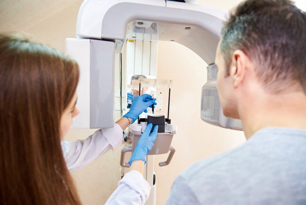
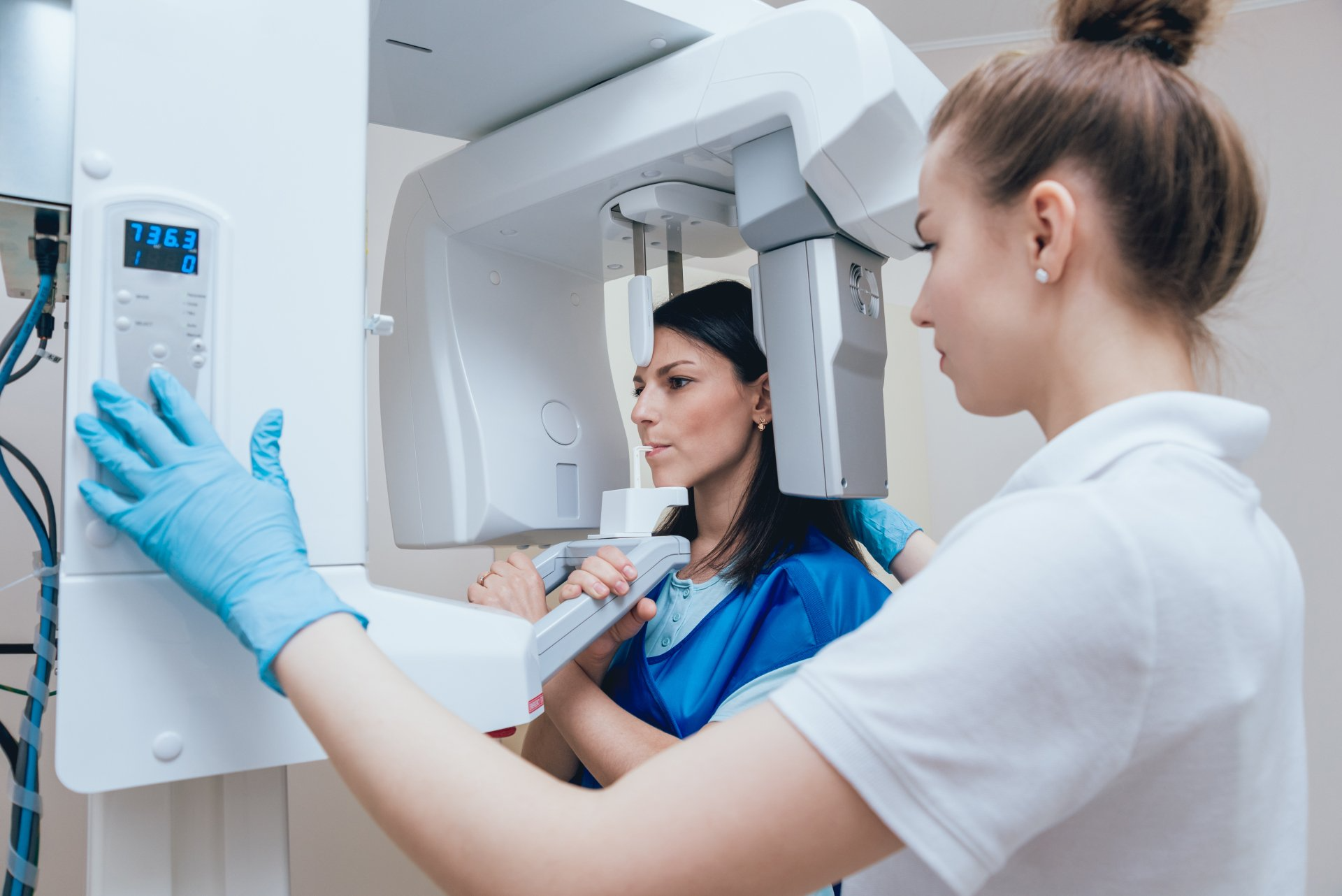
CBCT Machine
Dental cone beam computed tomography (CBCT) is a special type of x-ray machine that is implemented in scenarios where normal dental or facial x-rays are insufficient. This variation of the CT scanner employs a special type of technology to generate 3D images of dental structures, soft tissues, nerve paths, and bone in the craniofacial area, in one scan. These images allow for more specific treatment planning. The CBCT machine has an x-ray beam, in the shape of a cone, which moves around you to create a large number of high-quality images, or views. It was developed as a means to produce similar images to what a CT provides, though with a significantly smaller and less costly machine that could be situated in an outpatient office. Providing detailed images of the bone, the CBCT machine evaluates diseases of the jaw, dentition, bony structures of the face, sinuses, and nasal cavity. One shortcoming is that it does not provide the comprehensive diagnostic information available with conventional CT, especially in the analyzing of soft tissue structures such as muscles, glands, nerves, and lymph nodes. The CBCT machine can also be used for reconstructive surgery, cephalometric analysis, locating the origin of pathology, surgical planning for impacted teeth, diagnosing temporomandibular joint disorder (TMD), and for the accurate placement of dental implants.
-
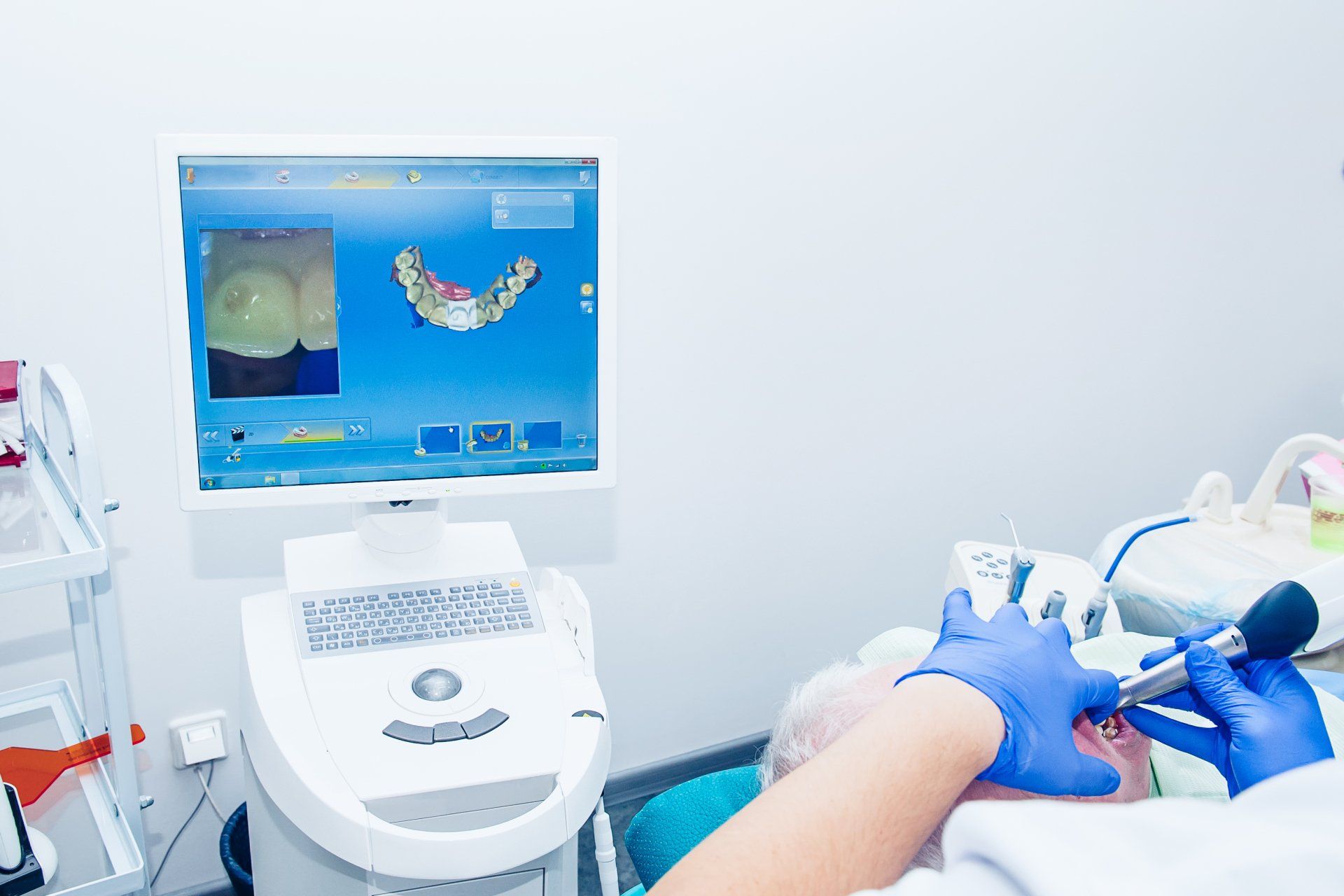
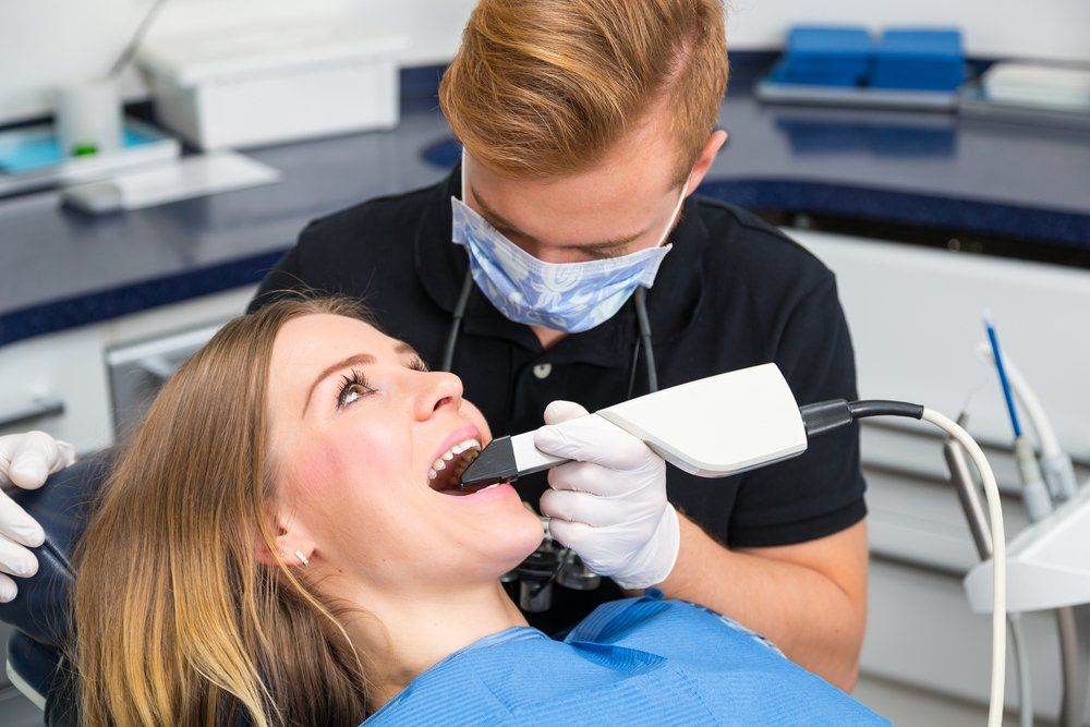
Intraoral Camera
About the same size as a marker, intraoral cameras are digital imaging tools used to create high-resolution images of your teeth, gums, and other hard-to-reach places in your mouth. Intraoral cameras help dental professionals detect dental issues, such as tooth decay, periodontal disease, and oral cancer. Other great benefits include:
- You can see, with precision, where you need to focus on brushing or flossing.
- You can see the difference before and after treatment.
- You can see magnified images of your teeth and gums, which helps dental professionals diagnose gum disease and cavities, and if caught early, can help prevent them.
- These photos provide proof for insurance companies to give you the coverage you need.
- Intraoral cameras also limit your time in the office because the images are produced in real-time, and the outcomes are available almost immediately.
-
Picasso Dental Laser
Diode Laser | The diode laser is useful for procedures that involve soft tissues, and are great for sterilizing endodontic canals, treating periodontal disease, and teeth whitening. It is a tool that offers a wide array of clinical treatment possibilities and is capable of great precision thanks to its portability and touchscreen controls. It has been shown to be helpful in treating challenging periodontal conditions while providing rapid healing and reduced swelling.
-
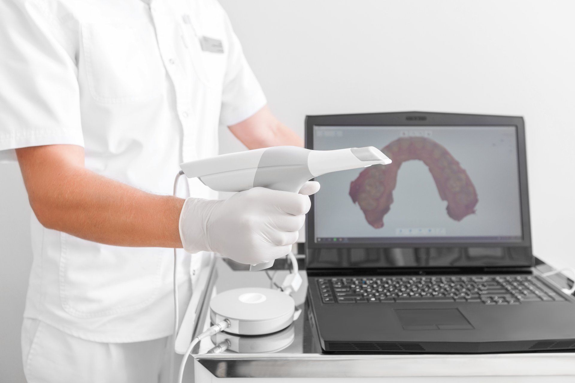
3Shape Digital Wand By Trios
Digital dentistry has taken its next step with the 3Shape Intraoral Trios Scanner, which is used to create a digital impression of a tooth's anatomy and the tri-dimensional position of dental implants. Information is electronically stored, and this form of digital dentistry affords patients the convenience of not having to sit through traditional impressions, with their unsavory tasting materials, bulky trays, and possible gagging effects. Around five percent of dentists worldwide currently use intraoral scanning but having the capabilities to make a replica of an entire mouth in less than two minutes, that statistic should only grow over time. Traditional methods of taking dental impressions, referred to as "analog", simply cannot keep up with the digital alternative. Digital imaging from the 3Shape Intraoral Trios Scanner is near flawless, an amazingly accurate tool for creating dental implants, crowns, and orthodontic aligner trays. As if that was not enough of an endorsement, the scanner also functions as a camera and operates at remarkable speeds.
-
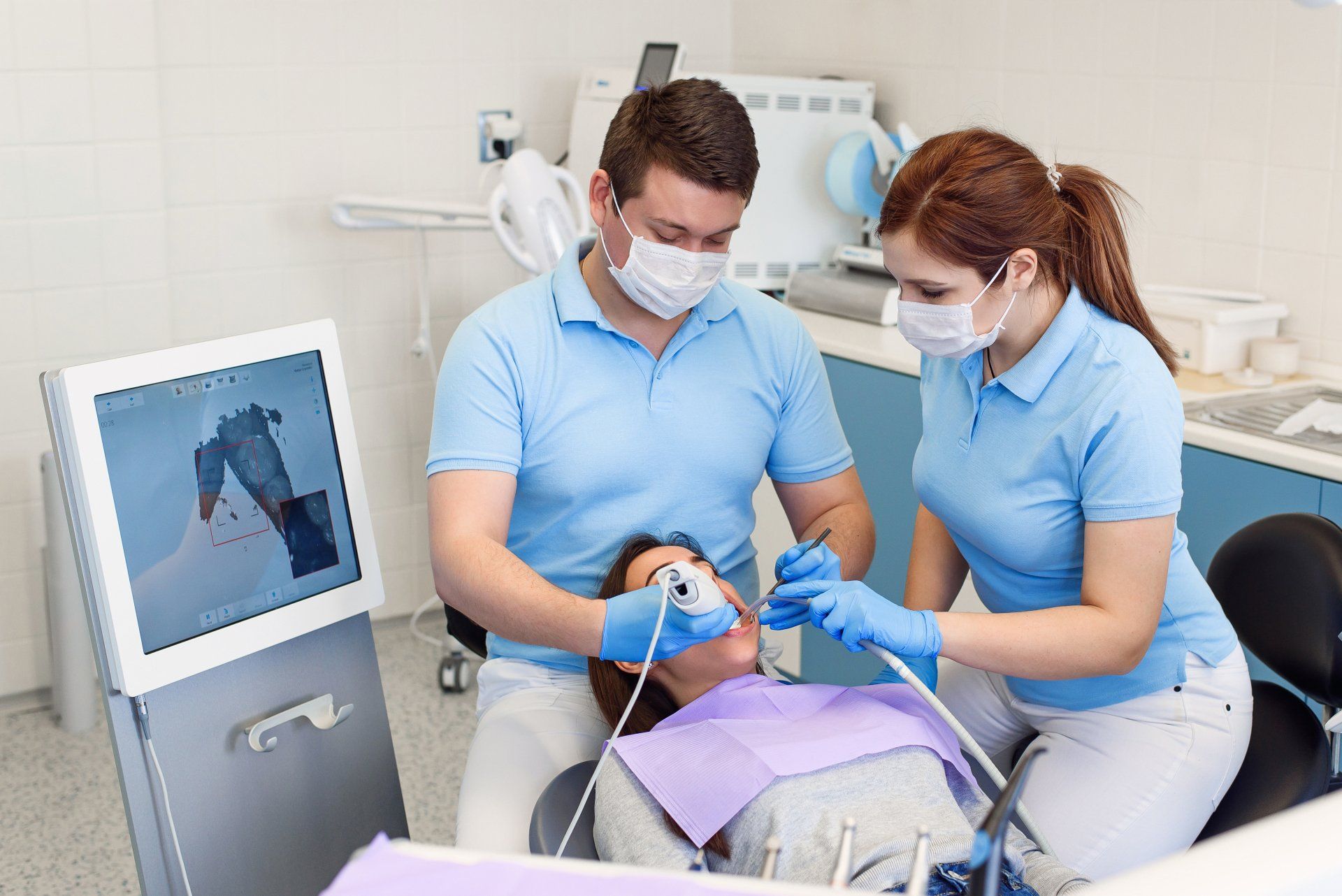
3D Intraoral Scanner
Our Intraoral Scanner is the newest form in digital imaging. It provides patients with a safe and complete oral impression without the use of radiation. The use of an Intraoral Scanner is also non-invasive and quite simple. The technology allows our patients to be in-and-out of the office in a few short minutes. This piece of equipment is environmentally friendly too, meaning no films, chemicals, or disposals of molds. We receive instant digital images of your full mouth which helps speed along your treatment plans. Experience the joys of living in the digital age.